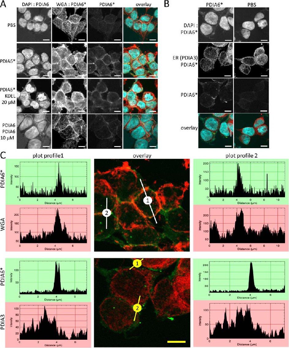
Fig. 3. (A) Fluorescently labeled PDIA6 (PDIA6*) associates with the cell surface of PDIA6. HEK293T cells were labeled for 3 hours with 3 µM PDIA6* alone or in the presence of either 20 µM KDEL peptide or 10 µM unlabeled PDIA6. Afterwards, the cells were stained with DAPI and WGA, and images were taken. (B) Fluorescently labeled PDIA6 does not travel back into the ER. After incubation of HEK293T cells with fluorescently labeled PDIA6, cells were permeabilized and stained with DAPI and against PDIA3 as a marker of the ER. No colocalization could be detected. (C) Plot profile of co-staining with fluorescent labeled PDIA6* either with WGA (cell surface) or PDIA3 (ER). The plot profile shows the intensities of pixels along the indicated lines in the corresponding images. X-axis represents the horizontal distance through the selection and the y-axis the vertically averaged pixel intensity. Although values of pixel intensities differ for each dye, maximum intensities could be detected in green (PDIA6*) and red (WGA) in similar pattern but not in case of co-detection of green (PDIA6*) and red (PDIA3). (A-C) Scale bars indicate 5 µm.